Learn more about our solutions.
Get echos read by top cardiologists assisted by AI.
Use our large collection of data for your project.
Learn more about our team of experts.
Read our latest news.
Reach out to us and integrate AI into your pipeline today!


Solutions
Using machine learning, we build tools for everything echo.
Take a tour of our solutions below.
iCardio.ai Ejection Fraction Simpson's Method
Ejection Fraction via Simpson's Method and other Left Ventricular Calcuations
Calculate left ventricular area and volume, ejection fraction, axis of symmetry, and diameters, all at systolic, diastolic and mid points in the cardiac cycle in the Apical, Two, Three, and Four Chamber perspectives.
iCardio.ai Ejection Fraction Teicholz's Method
Ejection Fraction via Teicholz's Method and Left Ventricular Calcuations
Ejection Fraction via a modified Teichholz's Method and Left Ventricular Calcuations at Mitral, Papillary and Apex levels in the Parasternal Short Axis perspective.
Left Atrium in Apical Views
Identification, segmentation and quantification of the left atrium.
Automatically determine left atrial function and dimensions.
Right Atrium in Apical Views
Identification, segmentation and quantification of the right atrium.
Automatically determine right atrial function and dimensions.
Aortic Annulus and Sinus of Valsalva in Parasternal Views
This model can automatically determine the diameter of at the aorta annulus. Our AI by default measures the area between the aorta annullus and the sinotubular junction, to extract additional area information and to reinforce conclusions behind the diamteric measurement of the sinotubular junction.
Sinotubular Junction in Parasternal Views
Automatically determine the diameter of the sinotubular junction across the cardiac and valve cycle and instantly flag abnormalities.
Left Ventricular Outflow Tract in the A5C Perspective
Quantify the area of the left ventricular outflow tract to produce a first-pass assessment.
Inferior Vena Cava
Quantification of the inferior vena cava from the right atrium to the hepatic vain.
Navigation and Guidance
This model extracts features in real-time using a mobilenet model architecture. By providing labeled features of the heart, ultrasound novices can orient themselves around the heart.
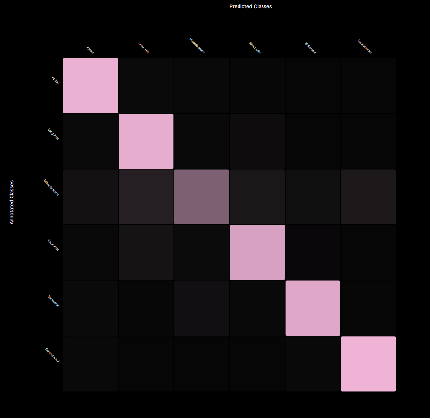
View Classification and Grading
This model extracts features in real-time using a mobilenet model architecture. By providing labeled features of the heart, ultrasound novices can orient themselves around the heart.
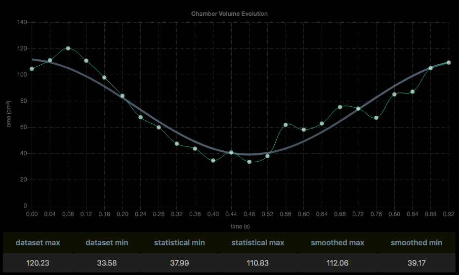
Performance and Data Science
All models undergo rigorous performance testing. In this example, we can visualize performance of the model's prediction over each frame of the cardiac cycle.